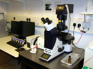
Leica TCS SPE inverted confocal microscope
...
| Section | |||||||||||||||
|---|---|---|---|---|---|---|---|---|---|---|---|---|---|---|---|
|
...
|
...
|
...
There is only one photomultiplier tube (PMT) so all multi-channel acquisition is done sequentially
A galvanometer driven stage insert is used to change the focus when optically sectioning samples. The speed of this device means that XZY scans are possible in addition to the conventional XYZ scan.
The ACS objective lenses, which have been designed to correct for colour registration errors caused by chromatic aberration, do not have a flat field, so best results are achieved at zooms ≥1.6.
There is also the option on this machine to carry out small screens thanks to a matrix screener software
The confocal scanning system has the following lasers:
Lasers and Detectors
- 405 nm (violet, for blue dyes like DAPI)
- 488 nm (blue, for green dyes like GFP)
- 561 nm (lime green, for red dyes like mCherry)
- 635 nm (red, for far red dyes like Cy5)
It has a quad dichroic mirror meaning a rapid switch between channels.
There is only one photomultiplier tube (PMT) so all multi-channel acquisition is done sequentially
A galvanometer driven stage insert is used to change the focus when optically sectioning samples. The speed of this device means that XZY scans are possible in addition to the conventional XYZ scan.
The ACS objective lenses, which have been designed to correct for colour registration errors caused by chromatic aberration, do not have a flat field, so best results are achieved at zooms ≥1.6.
There is also the option on this machine to carry out small screens thanks to a matrix screener software
The following PDF lists the recommended start-up and shut-down order for the machine: SPE3_StartUp_Shut Down.pdf
Changing the Galvo Z stage inserts
There are three inserts for the Galvo Z stage, although in practice you'll probably only need to use two at most. The main ones are the universal adjustable insert that will take slides, dishes, Lab-tek chambers etc and the multi-well plate insert. The universal insert should be fitted by default since it suits the most users. If you use the insert for the multi-well plate then you MUST replace the universal adjustable stage insert once you have finished. The screws for the universal insert are very small so you MUST be careful not to drop any into the microscope stand; particularly under the objective lens nose-piece. If a screw rolls under the nose-piece then contact a member of light microscope staff immediately.
Links:
...
|