Confocal Microscopy - An Introduction
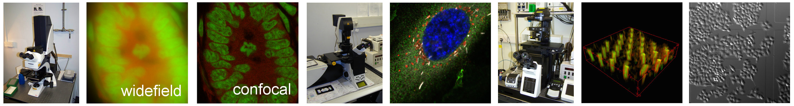
In wide-field microscopy the contrast of the focused image can sometimes be reduced by light from out of the focus plane - a phenomenon called blur. This is especially problematic for thick fluorescent specimens such as tissue or small organisms. Confocal microscopes are designed to reduce the amount of out-of-focus light reaching the detector.
Laser scanning confocal microscopes illuminate very small volumes within the sample with a focused laser spot so that the entire sample is not illuminated at the same time. The light emitted from the spot travels back through the microscope and through a pinhole in front of the detector. This pinhole is in a conjugate plane to the focused laser spot in the sample plane, so an image of the spot is formed at the pinhole. Light from above and below the plane of focus will come into focus above and below the pinhole plane and most will not go through the pinhole. So most of the light reaching the detector is from the focused spot.
Since a confocal image represents a single ‘slice’ from a three-dimensional sample with next to no light coming from adjacent slices, a motorised focus mechanism can be used to acquire every slice from the sample in sequence. These slices can then be reconstructed into a stack that represents the entire 3D structure. This is called optical sectioning.
Another technique called deconvolution can be used to computationally reassign light back to its correct plane of focus in both wide-field and confocal images by applying information known about how the microscope distorts the image of the relative to the object (the point spread function) to a reconstruction algorithm.
Confocal laser scanning microscopes
Most of the confocal microscopes in the LMCB use a standard design first developed in the 1980s in which a single laser beam is deflected off galvanometer driven mirrors and scanned across the specimen in a raster pattern. Most confocal microscopes still use this design for scanning the specimen - and if someone talks about a 'confocal microscope' then most of the time this is the kind of device they will be referring to. This design is usually referred to in technical and methods papers as a confocal laser scanning microscope (CLSM) although it would be less ambiguous to call it a single point confocal laser scanning microscope. The CLSM design allows for precise adjustment of the pinhole to reject out-of-focus light and free adjustment of the optical zoom to set the optimal pixel size for sampling. These factors mean that the CLSM gives the best resolution over the widest range of objective lens magnifications and numerical apertures of all the various confocal designs. It also makes the design extremely flexible. The main disadvantage is slow speed - since it takes a long time to scan a single laser spot over a specimen, especially at high resolution.
Spinning disc confocal microscopes
The Vox confocal microscope is a spinning disc confocal, also sometimes called a Nipkow disc confocal after the original image scanning device patented in 1884 by Paul Gottlieb Nipkow. The purpose of the spinning disc is to increase the speed of capture of optically sectioned images by illuminating multiple spots on the specimen in parallel rather than having a single laser spot.